


March 2026
In this VETgirl online veterinary continuing education blog, Dr. Joya Griffin, DACVD discusses allergic skin disease in cats! Diagnosing allergic skin disease in cats is more challenging than dogs because less is known about skin barrier function and clinical signs vary significantly! Complicating matters, the terminology for the condition is still debated but that doesn’t mean cats are not affected and deserve our attention. Read below to learn more!
The Answers You Really Want to Know About Feline Atopic Skin Syndrome
Dr. Joya Griffin, DACVD, Animal Dermatology Clinic, Louisville, KY
Far less is known about allergic skin disease in cats compared to dogs and humans. Feline patients often present differently, with variable clinical signs, inconsistent demonstration of IgE, and limited knowledge regarding their skin barrier function. There is debate as to whether atopic dermatitis (AD) even exists in the cat. For years, the term non-flea, non-food hypersensitivity dermatitis (NFNFHD) was the preferred term and more recently the term feline atopic skin syndrome (FASS) is often favored. FASS is one component of the broader feline atopic syndrome (FAS) which encompasses a spectrum of hypersensitivity disorders with varying presentations that affect the skin, gastrointestinal and respiratory tracts. Whatever the terminology chosen little is known about allergic dermatitis in cats relative to their canine counterparts. In one retrospective study, NFNFHD was found in 12% of allergic cats. The face and ventrum were most commonly affected and allergen-specific IgE was detected in almost 70% of these cats suggesting an allergic component. But overall, few studies exist.

Common facial presentation of feline allergic dermatitis. Photos courtesy of Dr. Joya Griffin, DACVD

Common ventral abdominal presentation of feline allergic dermatitis. Photo courtesy of Dr. Joya Griffin, DACVD
Likewise, little is known about the microbiota of allergic cats. Secondary bacterial infections are reported in less than 50% of cases which is less common than reported in dogs. This may be due to decreased corneocyte adherence of bacteria, though more recent studies show higher prevalence of infections. Cats with bacterial infections may present in varying ways that are less commonly seen in dogs. They can have lesions as subtle as erythema and seborrhea or present with classic pustules. Other signs of infection include crusted papules, eroded to ulcerated plaques, and linear to nodular granulomatous lesions that may be ulcerated. Because these presentations are similar in appearance to the reaction patterns that occur in allergic cats, secondary staphylococcal infections may go overlooked. A fungal dysbiosis was found in the skin of allergic cats in one study with Malassezia being isolated in higher numbers in affected sites pointing toward the importance of looking for and treating secondary infections in cats that present with skin disease.
The exact pathomechanism of FASS remains unclear, however, histopathologic studies show similar inflammatory reactions as found in the canine patient. T cell involvement is apparent but whether IL-31 is a key pruritogenic cytokine remains unclear.
Cats with allergic skin disease, whether related to food, fleas or environmental allergens, present with varying reaction patterns. FASS can also been seen in conjunction with allergic asthma, conjunctivitis, and rhinitis in some cases. The cutaneous reaction patterns in the cat include head and neck pruritus (or lesion-less pruritus), eosinophilic granuloma complex, self-induced hair loss (“fur-mowing”), papulocrustous dermatitis (“miliary dermatitis”). These reaction patterns do not indicate a specific hypersensitivity disorder. They can be seen alone or in various combinations in the same cat and can present differently from season to season or as allergy symptoms worsen over time. For example, one year the cat may have an indolent ulcer as a reaction pattern due to pollen allergens. The following year may erupt with miliary dermatitis. These reaction patterns can also occur in non-hypersensitivity diseases as well as atopic dermatitis, food hypersensitivity, insect hypersensitivity, adverse cutaneous drug reaction, contact hypersensitivity, and intestinal parasite hypersensitivity.
Feline Reaction Patterns:
- Head and neck pruritus. The pruritus may be focused around the head and neck but can also be generalized and is not pathognomonic for one type of hypersensitivity disorder. Differential diagnoses include atopic dermatitis, food hypersensitivity, adverse cutaneous drug reaction, flea allergy dermatitis.
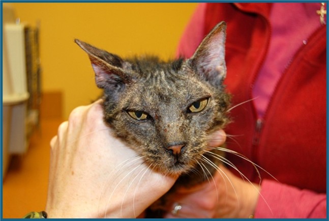
Facial pruritus in a cat. Photo courtesy of Dr. Joya Griffin, DACVD
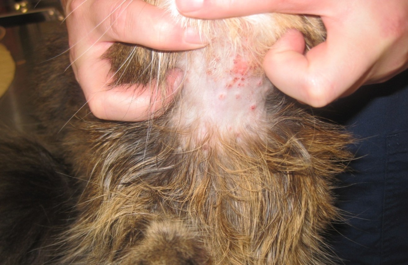
- Miliary dermatitis which appear as pinpoint crusted papules that are often rimmed by erythema and felt before they are seen as they can underly haired skin. Can be found commonly around the head and the neck and on the dorsum. Differentials include hypersensitivity disorders, bacterial infection. drug hypersensitivity, hypereosinophilic syndrome, ectoparasites: Cheyletiella, Otodectes, Lynxacarus, chiggers, lice, dermatophytosis.

Miliary dermatitis on a cat’s neck. Photo courtesy of Dr. Joya Griffin, DACVD
- Eosinophilic Granuloma Complex which includes the indolent ulcers, eosinophilic plaque, eosinophilic (linear) granuloma and eosinophilic granuloma causing a swollen or “pouty”-appearing chin. Differentials include hypersensitivity disorders or idiopathic and in the case of indolent ulcers can mimic squamous cell carcinoma. If not responding to treatment, biopsy is warranted.

Eosinophilic plaques on the ventral abdomen of a cat. Photo courtesy of Dr. Joya Griffin, DACVD
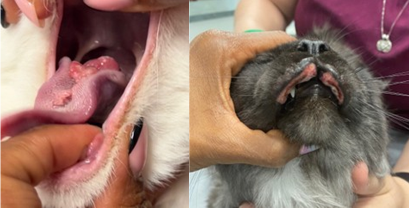
Eosinophilic granulomas on the tongue (left) and indolent ulcers on the lips (right) can be components of eosinophilic granuloma complex in cats. Photos courtesy of Dr. Joya Griffin, DACVD
- Self-induced hair loss and trauma, aka the “fur-mowing” cat (symmetric lesion-less pruritus). History is particularly important and can be challenging as some cats do not report excessive licking or grooming. Cats can often groom in private. Owners instead, may report excessive licking or grooming, vomiting of hairballs, or note hair in the feces, or large tufts of hair in the environment. On close examination, barbered or broken hairs can be seen. Can prove whether a cat is causing hair loss and it is not spontaneous by performing an “E-collar test” or by examining hairs via trichogram to see if hairs are broken or split from excessive grooming. Differential diagnoses include atopic dermatitis, food hypersensitivity, adverse cutaneous drug reaction, ectoparasites (fleas, Cheyletiella, Otodectes, Demodex gatoi), hyperthyroidism, and psychogenic (less likely if symmetric).
Diagnostic Approach to the Pruritic Cat:

Self-induced hair loss on the ventral abdomen of a cat. Photo courtesy of Dr. Joya Griffin, DACVD
Diagnosing FASS in the cat is a diagnosis of exclusion as it is in the dog. Biggest challenges come from difficulty doing diet trials in cats especially in multi-cat households or when owners cannot prevent the cat from roaming outdoors. Some feline owners are also insistent that their cat will not tolerate any diet change or is a “picky eater”. Ectoparasitic control can also be a difficult topic to approach when owners insist that their cat is indoor only and thus could not possibly be exposed to parasites or pollens for that matter. In fact, indoor air quality is almost identical to outdoor air quality so indoor only cats are not exempt from environmental allergies. Looking into the origin of the cat (likely at one point was outdoor, perhaps a stray), other animals that go outdoors and are in contact with the indoor cat, and new cats that have joined the household can serve as a risk factor for Demodex gatoi or fleas. Dermatophytosis should also be excluded as some cats with ringworm can be pruritic due to the cutaneous inflammatory response.
Treatment:
The approach to treatment in the allergic cat is similar to that of the dog, however, there are limited options for medical management of pruritus that are FDA approved for use. Antihistamines, glucocorticoids and cyclosporine are often used extra-labelly. Atopica® liquid is the only licensed medication for the treatment of FASS in cats and can offer good relief of symptoms but generally requires daily administration initially and has a lag period before improvement is seen that lasts 3-4 weeks. Once symptoms are controlled, tapering to every other day may be possible. Compounded cyclosporine in the form of flavored chewables, liquids or smaller capsules can be given to cats who are challenging to medicate.
Oclacitinib (Apoquel®) has shown efficacy in reducing clinical signs in a few limited studies in cats with FASS; however, is not labelled for use in cats and should be used with caution due to narrow margin of safety in cats. If used CBC and biochemistry analysis is warranted. Lokivetmab (Cytopoint®) is not recommended for use in cats due to the potential for severe adverse reactions to this foreign protein (caninized monoclonal antibody).
Allergen-specific immunotherapy remains my treatment of choice as a targeted, long-term, drug-free option. The goal of immunotherapy is to manage the symptoms without daily chronic medications that often carry side effects. The added benefit of not having to catch a cat daily for oral medications is often appealing and provides a better quality of life for the owner and the cat. Intradermal testing is performed to identify specific allergen reactivity, however, can be challenging due to transient and weak reactions. Serologic testing is frequently utilized either alone or in combination with intradermal testing. Studies on the efficacy of immunotherapy in cats are also limited, however, I have had good success with immunotherapy with decreased need for chronic medications and fewer allergic flares over time.
Subcutaneous immunotherapy is well-tolerated in most cats. Transdermal immunotherapy is a newer mode of administration and is very well-accepted by most felines and has shown good efficacy so far (Allibre Animal Inc.©). Sublingual immunotherapy is less often chosen due to the need for once to twice daily administration but is a good option for some owners. While beginning immunotherapy, low-dose alternate day glucocorticoids or Atopica® liquid is used to provide relief of pruritus. Antihistamines provide little relief alone in the majority of cats but may provide steroid-sparing effect in some cats.
Limited studies have shown mild to moderate efficacy to support use of Redonyl® and Cerenia®. Redonyl® and now Dermaquin® Skin Support Supplement contain palmitoylethanolamide (PEA), an anti-inflammatory endocannabinoid supplement that reduces mast cell degranulation. PEA has been shown to reduce pruritus and lesions in atopic dogs. In a small study, Redonyl® was able to maintain methylprednisolone-reduced level of pruritus and lesions than placebo. Maropitant (Cerenia®) reduced pruritus in 11/12 cats by more than 50% and 10/12 cats had reduction in lesions on the skin in one open study conducted over 4 weeks.
Topical therapy is important in cats to reduce allergen exposure and restore the barrier function of the skin. Key is to find the product that works best for the pet parent and is well-tolerated for the cat. Medicated wipes or mousse are useful to treat inflamed or focally infected areas, though some cats will tolerate bathing. I utilize low-potency steroids and tacrolimus for focally inflamed and pruritic lesions along with topical spot on products that contain essential oils and fatty acids like (Dermoscent® Essential 6 Spot-on, Atopivet® spot-on, mousse, or collar).
Abbreviations:
AD atopic dermatitis
FAS feline atopic syndrome
FASS feline atopic skin syndrome
NFNFHD non-flea, non-food hypersensitivity dermatitis
References:
- Miller WH, Griffin CE, Campbell, KL. Hypersensitivity Disorders. In: Muller and Kirk’s Small Animal Dermatology, 7th Edn, St. Louis: Eslevier, 2013:388-392, 402-405.
- Ravens PA, Xu BJ, Vogelnest LJ. Feline atopic dermatitis: a retrospective study of 45 cases (2001-2012). Vet Dermatol. 2014;25(2):95-102.
- Diesel A. Cutaneous hypersensitivity dermatoses in the feline patient: a review of allergic skin disease in cats. Vet Sci. 2017;4(2):25.
- Marsella R, De Benedetto A. Atopic dermatitis in animals and people: an update and comparative review. Vet Sci. 2017;4(3):37.
- Noli C, Matricoti I, Schievano C. A double-blinded, randomized, methylprednisolone-controlled study on the efficacy of oclacitinib in the management of pruritus in cats with nonflea nonfood-induced hypersensitivity dermatitis. Vet Dermatol. 2019;30(2):110-e30.
- Santoro D, Pucheu-Haston CM, Prost C, et al. Clinical signs and diagnosis of feline atopic syndrome: detailed guidelines for a correct diagnosis. Vet Dermatol. 2021;32(1):26-40.
- Halliwell R, Pucheu-Haston CM, Olivry T, et al. Feline allergic diseases: introduction and proposed nomenclature. Vet Dermatol. 2021;32:8-12.
- Mueller RS, Nuttall T, Prost C, et al. Treatment of the feline atopic syndrome- a systematic review. Vet Dermatol. 2021;32:43-60.
- Maina E, Fontaine J. Use of maropitant for the control of pruritus in non-flea, non-food-induced feline hypersensitivity dermatitis: an open-label, uncontrolled pilot study. J Feline Med Surg. 2019;21(10):967–972.
- Scarampella F, Abramo F, Noli C. Clinical and histological evaluation of an analogue of palmitoylethanolamide, PLR 120 (comicronized Palmidrol INN) in cats with eosinophilic granuloma and eosinophilic plaque: a pilot study. Vet 2001;12:29–39.


Only VETgirl members can leave comments. Sign In or Join VETgirl now!